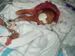
Entry for March 21, 2006
Went to Caitlin's eye dr today and she has stage 3 of ROP ,She is going to have laser surgery on Thursday so please pray it goes well. She has been such a good girl today and once again daddy has not let her go lol. We have her peditrician apt tomorrow so we will see how her weight is. Today I ended up waiting in the lobby for Caitlin's apt because there were alot of people in the actual office and so many people asked to see her and I said no to everyone. I felt so rude but hey I have to protect my baby from germs!!! It was funny to see some of the reactions to my awnser but I wasn't there to please them. lol . Well here is a bigt explantion of ROP again.
XI. RETINOPATHY OF PREMATURITY
Retinopathy of Prematurity (ROP) causes a disease of the retina. It affects prematurely born babies. It consists of abnormal retinal vessels that grow mostly in an area where normal vessels have not yet grown in the retina (Fig. 20). ROP is divided into stages 1 to 5. Stages 1 and 2 do not usually require treatment. Some babies who have develop stage 3 ROP require treatment. The treatment is usually performed either by laser or cryotherapy (freezing). Laser is more commonly used now than cryotherapy because of various advances in the laser treatment. The ROP in stage 3 that requires treatment is generally called threshold disease. The majority (95%) of the babies who require laser or cryotherapy develop threshold disease between 32 and 42 weeks after conception. The post-conceptional age is calculated from the presumed day of conception.
The area of the retina affected by ROP is divided into three zones (Fig. 21). Zone 1 is the most centrally located, and ROP develops in this zone if the retina in this area is most underdeveloped. Disease in zone 1 is more severe compared with disease limited to zones 2 or 3. Timing is one of the important factors that make the treatment successful in ROP, because the disease can advance very quickly and delayed treatment often reduces the chances of success. The rapidly progressing ROP is called Rush disease, and it is usually associated with very extensive or aggressive growth of abnormal blood vessels. Abnormal dilatation of retinal veins with florid abnormal new vessels is called Plus disease.
Zone 1 is the most posterior retina, that contains the optic nerve and the macula (zone of acute vision). Zone 2 is the intermediate zone where blood vessels often stop in ROP. Zone 3 is the peripheral zone of the retina, where vessels are absent in ROP, but present in normal eyes.
The treatment’s goal is to destroy the retina that is deprived of retinal vessels. This helps to shrink the new vessels and prevents the formation of dense scars that usually follow. The dense scars cause traction on the retina. The result is distortion of the normal orientation of the retina and impairment of vision. In other cases, the retina detaches from the wall of the eye. The treatment extinguishes the "fire" of growing abnormal vessels in the eye, ultimately preventing the retina from detaching or from being severely distorted. Retinal detachment is the main cause of total blindness in ROP. Contrary to what happens in adults, retinal detachment in premature babies is generally caused by the traction of scar tissue on the retinal periphery. Small breaks in the peripheral retina are relatively rare in ROP, and a macular break is exceptional in this type of case. Though many babies have been saved from blindness. Though many babies have been saved from blindness by laser or cryotherapy the treatment is not always successful. ROP in some babies continues to progress to stage 4, (partial retinal detachment) and stage 5, (total retinal detachment).
Retinal Detachment In ROP
Retinal detachment is a serious consequence of stages 4 and 5 ROP. The scar produced by abnormal vessels inside the eye is the usual cause. The scar pulls the retina away from the wall of the eye. This scarring is not the result of laser or cryotherapy.
Stage 4 ROP causes partial retinal detachment. At this stage, the retina may sometimes reattach spontaneously. Retinal detachment which is unlikely to regress requires treatment. Treatment modalities are cryopexy for shallow retinal detachment, or scleral buckling for more advanced cases. The latter consists of using a silicone belt around the globe. In other cases, a vitrectomy operation is indicated. Stage 4 may progress quickly in some babies and its treatment has frequently been unsuccessful leading to stage 5 ROP.
Stage 5 ROP shows a total retinal detachment and is the end stage of the disease. In its most severe form, stage 5 shows a dense white membrane or scar behind the lens of the eye (Fig. 22). The detached retina is adherent to this scar tissue. The condition was previously called retrolental fibroplasia or RLF, because the scar is white and visible through the pupil, the condition is sometimes called leukocoria or white pupil. Spontaneous reattachment of the retina rarely happens in stage 5 ROP. If the eye is left alone at this stage, the baby becomes permanently and often totally blind in both eyes. In addition, some babies develop high intraocular pressure, causing glaucoma. Others develop problems leading to clouding of the cornea (front part of the eye).
From the late 70’s through the early 80’s, the clinical scientists headed by Dr. Charles Schepens developed a new surgical technique called open-sky vitrectomy to treat severe retinal detachment in general. The open-sky vitrectomy turned out to be quite effective in the most severe type of ROP, namely stage 5. Experience has shown that this condition cannot be treated successfully by any other method of surgery. Using this technique, Dr. Tatsuo Hirose has operated upon more than 1200 cases of advanced stage 5 ROP, many of which had been considered hopeless elsewhere. The results are encouraging. They vary depending upon the condition of the eye, the patient’s age, and other factors. Poor postoperative vision, in patients affected by advanced stage 5 ROP, is mainly due to the fact that the retina of those patients is incompletely developed.
The baby with stage 5 ROP who recovered some sight post operatively enters the world of the seeing from a world of the totally blind. This world is totally new to the baby, in spite of his incomplete visual recovery. Although the baby may only perceive vaguely the form and color of objects he/she is quick to exploit this partial ability with remarkable benefit.
Observation. Linda (not her real name) was a premature baby aged 6 months when she came from a central European country for treatment. She suffered from a total retinal detachment in both eyes, with a bilateral white pupil. After an open-sky operation in the most promising eye, she recovered 20/600 vision. At age seven, she learned to ride a bicycle and never had an accident in spite of her markedly diminished vision. Her continued progress required constant care by the parents postoperatively. The reason is that the child’s recovering retina has the ability to improve further, after a successful operation. With training, such babies often learn to distinguish several colors.
This case illustrates that babies who never had vision, like those affected by advanced stage 5 ROP, become able to exploit their incomplete postoperative visual recovery with astonishing agility. They are able to navigate between obstacles much better than would adults with the same diminished vision. The anatomical reattachment of the retina after open-sky vitrectomy, in the advanced stage 5 ROP, is 40%. Prior to the use of the open-sky operation, it was not above 6%. The best-corrected visual acuity measured by special procedures in children who had a white pupil, after open-sky vitrectomy, is 20/200. A majority of patients developed vision of less than 20/200, but this ambulatory vision was sufficient for them to navigate and recognize large objects. A small percentage of babies operated upon never developed useful vision, even after the retina had been completely reattached.
The optimal age for open-sky vitrectomy, in infants with advanced stage 5 ROP, is usually between 6 months and 1 year. There are some exceptions. Babies whose ROP is inactive, completely “dry,” without any neovascularization, may be operated upon before they reach six months of age. However, it is difficult to judge whether or not the ROP is inactive in an eye whose retina is not visible. In patients with advanced stage 5 ROP and whose intraocular abnormal blood vessels are not extensive, a less invasive operation such as a scleral buckling procedure, or a closed vitrectomy may be tried, even though the baby is less than 6 months of age. Babies whose eyes show massive subretinal hemorrhage, demonstrated by ultrasound, and babies with severe general physical abnormalities such as marked brain damage will not benefit from any type of eye surgery. However, many babies are abandoned simply because they have developed an advanced stage 5 ROP, in spite of the fact that open-sky vitrectomy still holds the hope they may see. The following examples are cases in which the open-sky vitrectomy was helpful in developing vision in babies with advanced stage 5 ROP.
Subscribe to:
Post Comments (Atom)


No comments:
Post a Comment